What causes it?
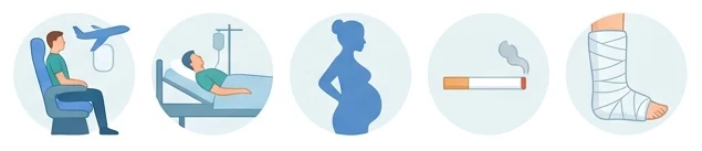
Clots form more easily when three conditions come together: slowing of the blood flow, damage to the vein wall, and a tendency of the blood to clot. In practice, the most common situations are:
- After surgery and prolonged bed rest
- Plaster casts, fractures, leg injuries
- Long flights, bus or car journeys
- Cancer and chemotherapy
- Pregnancy, the postpartum period, contraceptive pills or hormone therapy
- Inherited clotting disorders and a family history of clots
- Older age, obesity, smoking
- A previous deep vein thrombosis
How does a clot form and progress?
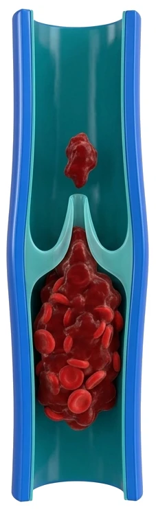 1
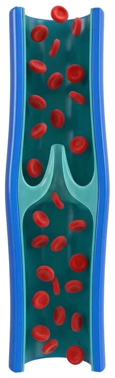
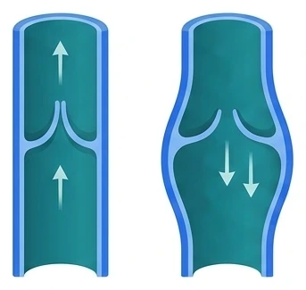
11. Normal flow
The valve leaflets open and close, and blood flows regularly toward the heart.
- Blood cells move freely and are dispersed within the vein
- Blood does not stand still in the valve pockets
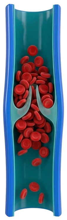
2. Slowing of flow
When immobile, blood slows down and pools in the valve pockets.
- Long journeys, bed rest, plaster casts
- The ground for a clot is prepared here
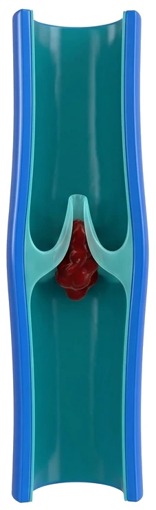
3. Clot begins
A small clot forms in a valve pocket.
- There are often no symptoms at this stage
- Prevention is therefore important for people at risk
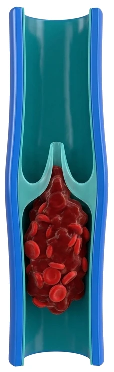
4. Vein blockage
The clot grows and fills the vein, and blood cannot flow back.
- Swelling, pain, redness and rising warmth in one leg
- Diagnosis is made by Doppler ultrasound and treatment starts immediately
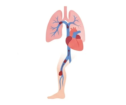
5. Clot breaks off
A piece of the clot can break off and travel to the heart and lungs (pulmonary embolism).
- Sudden shortness of breath, chest pain, palpitations
- Life-threatening: call 112 without delay

What are the signs of a clot in the leg?
A clot in the leg (deep vein thrombosis) often shows up with the symptoms below. Symptoms may be mild; some progress without any complaints.
- Swelling that increases over days, usually in one leg
- Tightness in the calf, cramp-like pain, tenderness when pressed
- Redness, a purplish colour change and rising warmth of the skin
- A feeling of fullness in the leg, and superficial veins becoming more prominent
- Sometimes the first sign, without any leg symptoms, is sudden shortness of breath or chest pain
This picture can be confused with varicose veins, but its course is different. Varicose swelling develops over years, increases in the evening and goes away in the morning. In deep vein thrombosis, the swelling appears within days and does not go away with rest.
Sudden swelling in one leg should not wait.
Diagnosis
After examination, the first step is imaging (Doppler ultrasound, CT/MR venography). It is painless and needle-free; it directly shows whether the vein is open, and the location and extent of the clot. If needed, a D-dimer blood test is used, and in selected patients a CT venography. In recurrent clots or clots occurring at a young age, inherited clotting disorders are investigated.
Treatment
Blood-thinning treatment
This is the foundation of treatment. The aim is to prevent the existing clot from growing and to prevent a new clot from forming and travelling to the lungs. The body gradually dissolves the clot over time. The medication and duration are determined by the location of the clot, its cause and the risk of recurrence. Stopping the medication on your own is the most dangerous behaviour in terms of the clot coming back.
Compression stockings
They reduce swelling and pain, and help lower the long-term risk of post-thrombotic syndrome. The correct compression class is determined by a physician.
Mechanical clot removal (thrombectomy)
In selected patients, especially when the clot involves the groin and the large veins above it, the leg is markedly swollen, and symptoms have just started, the clot is removed through a catheter. No incision is made. The aim is to open the vein early, reduce pressure in the leg, and reduce valve damage and therefore lasting swelling that could develop later. This procedure is not suitable for every patient; the decision is based on imaging and the clinical picture.
Vena cava filter
In a limited number of patients who cannot take blood thinners, or who continue to throw clots despite treatment, a filter that traps clots can be placed in the main vein.
What happens afterwards?
In some patients, lasting leg swelling, a feeling of heaviness, skin colour changes and, rarely, ulcers can develop months or years later. This is called post-thrombotic syndrome. The three main things that reduce the risk are: starting treatment early, taking the blood thinner regularly for the recommended duration, and not skipping compression stockings.
Proportion of people with a leg clot who develop a lasting leg complaint within 2 years
%0
Despite blood-thinning treatment.
This is called post-thrombotic syndrome.
In a randomised study of 692 patients with clots in the deep veins above and behind the knee, the group receiving only blood-thinning treatment.
Vedantham S et al. Pharmacomechanical catheter-directed thrombolysis for deep-vein thrombosis.
N Engl J Med 2017;377(23):2240-2252. doi:10.1056/NEJMoa1615066
Prevention
- On long journeys, stand up and walk every hour, and do ankle movements while seated
- Drink enough fluids
- Move early after surgery, to the extent your physician allows
- Do not skip the protective treatment recommended for high-risk procedures
- Stop smoking and control your weight
Frequently asked questions
What does 'a clot travelling to the leg' mean?
What people call a clot 'going to the leg' is most often deep vein thrombosis: a clot forming in the deep veins of the leg. Less commonly, a clot that breaks off from the heart and blocks an artery of the leg is also described this way; in that case the leg suddenly becomes cold, pale and very painful. Both situations are emergencies and should be assessed without delay.
Is the swelling in my leg from varicose veins or a clot?
Timing is what distinguishes them. Varicose swelling develops over years, increases in the evening, goes away in the morning and is usually on both sides. Swelling caused by a clot starts within days, affects one leg and does not go away with rest. The definitive distinction is made by imaging.
How long will I need to take a blood thinner?
The duration varies from person to person. For a first clot caused by a temporary cause, a few months of treatment is usually enough. The duration is extended when no cause is found, when clots recur, or when a lasting risk such as cancer persists. The treating physician makes this decision.
Can I walk, or do I need to stay in bed?
In the past, strict bed rest was recommended, but this approach has now been abandoned. Once treatment has started, walking with compression stockings reduces swelling and pain. Strenuous sports are avoided at the beginning.
Does it come back?
There is a risk of recurrence, and it depends on the cause. Clots caused by a temporary trigger (surgery, a plaster cast, a long journey) carry a lower risk. Clots that occur spontaneously carry a higher risk; treatment duration and follow-up are planned differently for these patients.
I was told a clot has travelled to my leg. What does that mean?
What people call a clot 'travelling' is the formation of a clot inside a deep vein. The real danger is that a piece of this clot breaks off and travels to the lungs. For this reason, treatment is started without losing time.
My leg swelled after a long journey. Is it dangerous?
Sitting still for long periods increases the risk of clots. Swelling, tightness and pain that appear in one leg after a journey should be taken seriously and assessed with imaging.
Examination and appointments. Patients from Bitlis, Tatvan, Van, Muş and the surrounding area are examined at the Cardiac and Vascular Surgery Outpatient Clinic of Tatvan State Hospital. Appointments are made through MHRS (Türkiye's national hospital appointment system); details are on the Appointments and Contact page.
Related pages
 Varicose vein treatmentSwelling that develops over years and increases in the evening
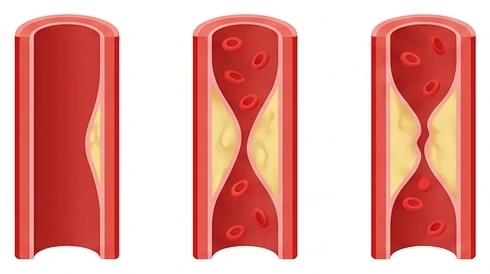 Peripheral artery diseaseA shortening walking distance
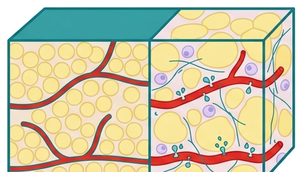 LipedemaSymmetrical, painful thickening of both legs
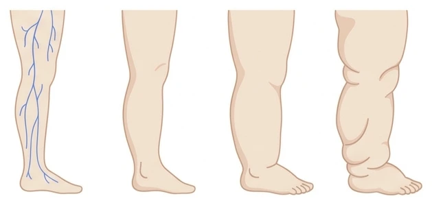 LymphedemaPersistent swelling that also involves the top of the foot